Introduction
Tuberculosis is a granulomatous, caseous necrotizing inflammatory disease caused by Mycobacterium tuberculosis [1]. Globally, tuberculosis is the leading infectious cause of death amongst adults [2]. Pulmonary tuberculosis is the most common type. Signs and symptoms of pulmonary tuberculosis typically include, but are not limited to, dry cough, hemoptysis, fever, shortness of breath, weight loss and night sweats [3]. In 2018, the WHO [4] reported 10 million novel tuberculosis cases, of which approximately 90% of affected individuals originated from 30 endemic countries [4]. Further, only approximately 70% of those novel cases were notified [4].
Pulmonary tuberculosis is a well-known entity. Extra pulmonary tuberculosis can occur up to 10% of pulmonary tuberculosis patients and may involve various sites of the body including skin, lymph nodes, pleura, bones and joints and meninges [5]. Urogenital tuberculosis accounts for 30 to 40% of the total extra pulmonary tuberculosis burden [5]. It spreads from the primary pulmonary focus via hematogenous route to the kidney and then to the ureters, bladder and urethra [5]. Due to insidious disease evolution with long latent period of average 22 years between the early infection to clinical urogenital tuberculosis, there is often a delay in diagnosis and treatment. Renal infections are usually asymptomatic but highly destructive, resulting in unilateral and rarely bilateral loss of renal function and renal failure on diagnosis [6]. Moreover, urogenital tuberculosis can be difficult to diagnose especially in communities that are not endemic for tuberculosis, leading to treatment delays. As migration continues in an increasingly globalized world, it is important that health care providers recognize the signs and symptoms of urogenital tuberculosis, especially among the urogynecological population where urinary investigations are often routine. Pharmacotherapy is the mainstay of the urogenital tuberculosis treatment. However, more than 50% of the patients may still need renal ablative surgery [7,8].
Renal tuberculosis presents a diagnostic challenge as it presents similarly to cystitis with lower urinary tract symptoms such as frequency, nocturia, hematuria or pyuria [9]. However, renal tuberculosis fails to respond to the usual empirical antibiotic regime [10] and majority yielding a negative culture on routine media even in the presence of pyuria [11]. Hence, it is always important to consider renal tuberculosis as a differential diagnosis in such a clinical presentation, especially since tuberculosis is endemic in Singapore.
Case report
A 63-year-old Chinese lady presented to the Urogynaecology clinic complaining of urinary frequency, nocturia and dysuria since few years. She did not have symptoms of urgency urinary incontinence or voiding dysfunction. She had no history of fever, weight or appetite loss and no history of pulmonary tuberculosis or any exposure to close contacts with tuberculosis. Her clinical and gynaecological examinations were unremarkable apart from atrophic changes consistent with atrophic vaginitis. She was advised on fluid modification including restriction of fluid intake up to 1.5 liter per day and reduction of caffeine intake. Physiotherapist referral for bladder training was also made. In addition, she was prescribed imipramine for her nocturia and vaginal estrogen cream was prescribed for her atrophic vaginitis.
Mid-stream urine microscopy and culture test was ordered to rule out a urinary tract infection. Urinalysis revealed microscopic haematuria (90/uL) and pyuria (1,080/uL), but urine culture was negative. Mid-stream urine test was repeated six weeks later, yielding persistent microscopic haematuria and pyuria with negative urine culture.
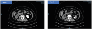
The serum creatinine came back normal. A computed tomographic urogram was performed which revealed calcifications in the left lower renal pole calyces with overlying cortical atrophy and scarring. There was also dilatation of the left upper renal pole calyces, possible urothelial thickening of the segmental infundibulum, left renal hilar lymphadenopathy and hypoperfusion of the renal parenchyma in the left upper renal pole (Figures 1 and 2). Overall, these features were suggestive of a chronic infectious or inflammatory process. A urine sample was sent off for urine acid-fast bacillus smear culture which returned positive for pan-sensitive Mycobacterium tuberculosis. She was advised to stop imipramine and was subsequently referred to the renal and infectious disease specialists for co-management. She was treated with isoniazid, rifampicin, ethambutol, pyrazinamide for 8 weeks and then continued with isoniazid and rifampicin for the next 9 weeks. Following completion of the tuberculosis treatment regime, she was well, and all her urinary symptoms including microscopic hematuria and pyuria were resolved.
Discussion
Renal tuberculosis is easily missed as most patients generally present with lower urinary symptoms similar to that of a simple cystitis or overactive bladder [7]. This often results in delayed diagnosis and may only be diagnosed after extensive renal damage has occurred. Renal tuberculosis usually occurs via hematogenous spread which could take place at the time of pulmonary infection or years later as a sequalae of reactivation after a long period of dormancy. It can be either a part of a disseminated infection or a localized genitourinary lesion [12]. The offending bacilli usually form caseating granulomas in the medullary interstitium, the site of preference in the kidney, causing local tissue destruction before involving the cortico-medullary junction. Other sequela of renal tuberculosis includes papillary necrosis where the infection causes vascular insufficiency of papillae vessels, ulceration of the calyces, involvement of the renal pelvis leading to tuberculous pyelonephritis or pyonephrosis. Scarring may develop within the renal pelvis with calcification found in 24%, identifiable as renal or ureteric stones in up to 19% of cases [8]. Other computed tomographic findings include echogenic kidneys, urothelial thickening, enlarged lymph nodes and fluid collections/abscesses on the native kidney [13]. Infection often spreads down the ureter into the bladder, producing mucosal and mural granulomatous lesions associated with scarring. Ureteric involvement may also produce irregular ureteric strictures and segmental dilation, leading to obstruction and reflux. Chronic tuberculosis is sometimes complicated by secondary amyloidosis [14]. There can also be renal parenchymal damage i.e. interstitial nephritis although rare [15]. Unilateral kidney involvement is also generally more common than bilateral involvement [6]. In a disseminated infection, blood-borne military tubercles are seen in the kidney globally, most notably the cortex though renal function is usually not compromised [16].
Treatment of renal tuberculosis generally consists of the usual anti-tuberculous drugs — first line being rifampicin, pyrazinamide, ethambutol, and isoniazid. Patients should be on an initial two-month phase of the four mentioned drugs, followed by a four-month continuation phase where rifampicin and isoniazid are administered. However in severe cases or in patients with immunosuppression, longer duration of therapy may be required [7]. Surgical intervention such as a stent or percutaneous nephrostomy can be considered in patients who develop ureteric strictures. A nephrectomy may be necessitated in cases with extensive renal damage [7,8].
Conclusion
In summary, this case highlights the possibility of underestimation and delayed diagnosis of renal tuberculosis as it usually presents with non-specific symptoms typical of a simple cystitis or overactive bladder. Hence, it is important to have increased diagnostic awareness and consider the differential diagnosis of renal tuberculosis so that prompt treatment can be initiated before the disease progresses to chronic renal failure.
Funding
None.
Disclosure of all authors:
No disclosures.
Conflicts of interest
The authors declare having no conflicts of interest.
Author contribution to the manuscript:
Kazila Bhutia and Han How Chuan: Patient care, writing of the manuscript, approval of the final version. Ng Ding Yi and Tanaka Dune: Writing of the manuscript, approval of the final version.
Consent
Written informed consent was obtained from the patient for publication of this case report and any accompanying images.